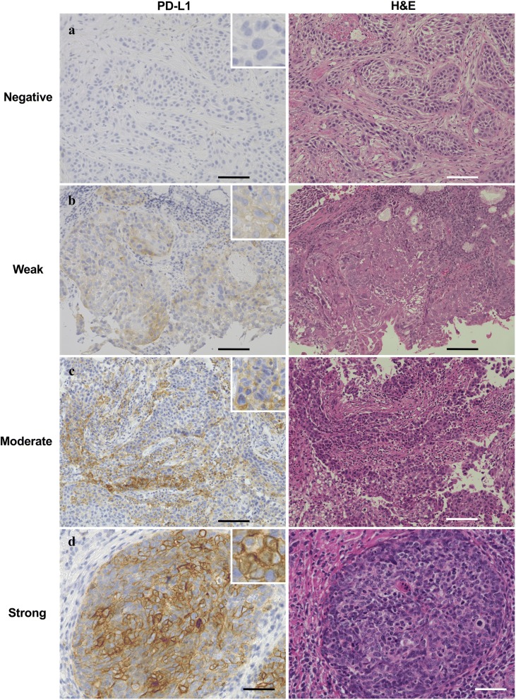
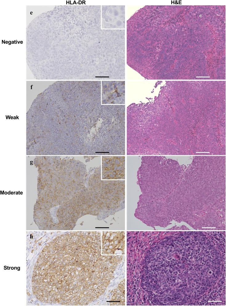
Fig. 1.
Expression levels of PD-L1 and HLA-DR in oropharynx squamous cell carcinoma tissue. Tissue specimens of patient with oropharynx squamous cell carcinoma were classified into four groups by immunostaining intensity for PD-L1 or HLA-DR: a, e negative staining; b, f weak staining; c, g moderate staining; d, h strong staining. H&E staining was shown in the right. Representative images are shown. Scale bar is 100 μm


