Figure 1.
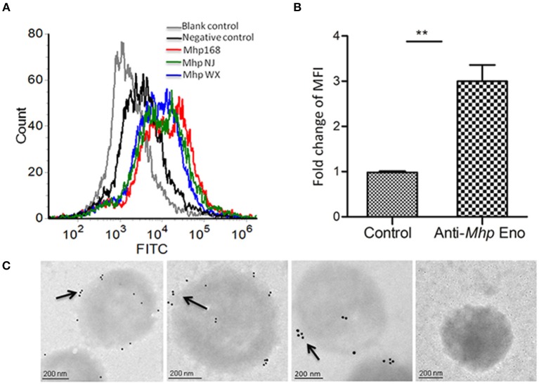
Detection of Mhp Eno on the surface of M. hyopneumoniae by flow cytometry and immune electron microscopy. (A) Blank control, M. hyopneumoniae strain 168 treated with PBS; negative control, M. hyopneumoniae strain 168 treated with preimmune serum; M. hyopneumoniae strains 168, NJ, and WX treated with anti-Mhp Eno serum. (B) The MFI level of M. hyopneumoniae incubated with anti-Mhp Eno sera is expressed as the percentage of that found for the corresponding strain incubated with preimmune sera. The asterisks above the charts indicate statistically significant differences. (C) From left to right, the first three M. hyopneumoniae strains, 168, NJ and WX, were treated with anti-Mhp Eno serum and secondary gold-conjugated antibodies. The last column indicates the M. hyopneumoniae strain treated with preimmune serum and secondary gold-conjugated antibodies.

