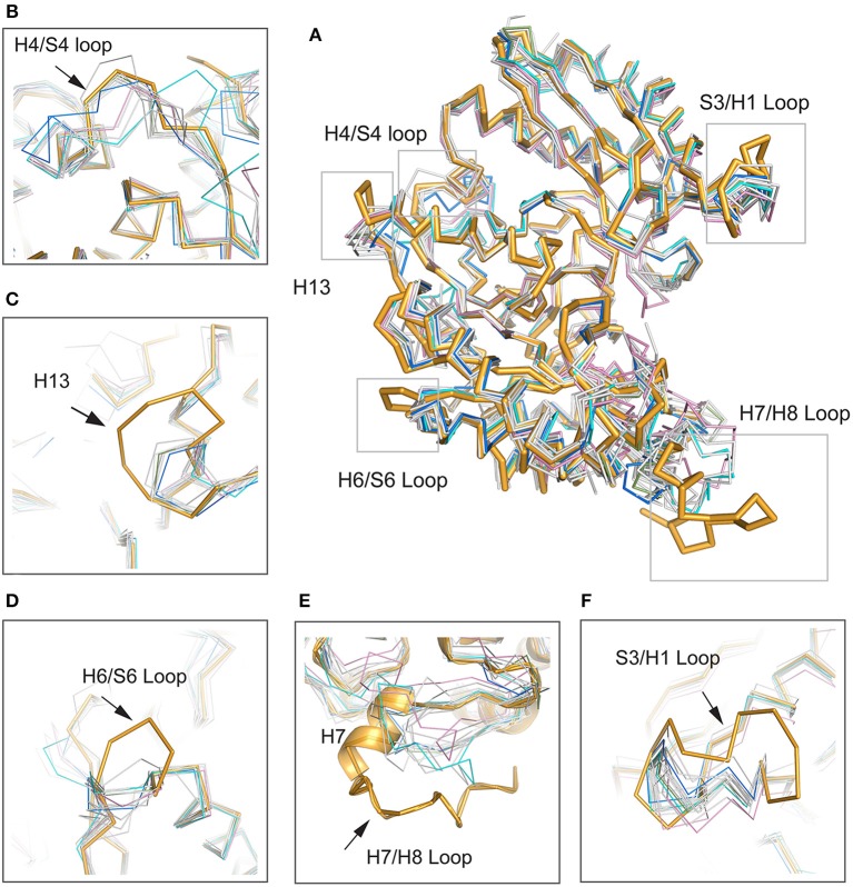
Figure 5.
Structural comparisons between Mhp Eno and other enolases. Mhp Eno is shown in bright orange. Enolases from human, yeast, E. coli and Bacillus subtilis are shown in pink, cyan, blue and green, respectively, and the other enolases are shown in white. The overall structure is shown in (A). (B–F) show enlarged pictures of the H4/S4 loop, H13, H6/S6 loop, H7/H8 loop, and S3/H1 loop regions. H7 is shown in both cartoon and ribbon forms in (E).

