
Figure 1.
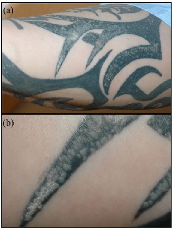
(a) Clinical photograph of the right ventral forearm showing white to skin-coloured flat-topped papules confined to areas of tattoo pigment. (b) Close-up of flat-topped papules with area of scale that developed after treatment with liquid nitrogen cryotherapy.