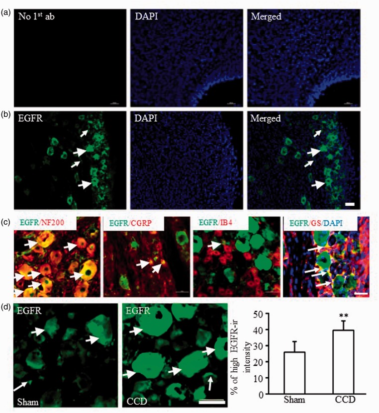
Figure 2.
Distribution of EGFR protein in the lumbar DRGs of rats. (a) No EGFR signals were detected in the control DRG slices without EGFR antibody incubation. (b) EGFR was expressed in the cytosol of neurons and nonneuronal cells around cellular nuclei (labeled by DAPI) in the DRG. (c) EGFR was colocalized with NF200, CGRP, IB4, or glutamine synthetase. (d) CCD increased the numbers of DRG neurons with high EGFR-ir intensity in the ipsilateral L4/L5 DRGs. N = 5 rats/group. **P < 0.01 versus the corresponding sham group by two-tailed unpaired Student’s t test. Short arrows: DRG neurons with high EGFR-ir intensity; Long arrows: glial cells with EGFR-ir. Scale bars: 50 µm. CCD: chronic compression of dorsal root ganglion; CGRP: calcitonin gene-related peptide; DAPI: 4′,6-diamidino-2-phenylindole; EGFR: epidermal growth factor receptor; EGFR-ir: EGFR immunoreactivity; IB4: isolectin B4; NF200, neurofilament-200.

