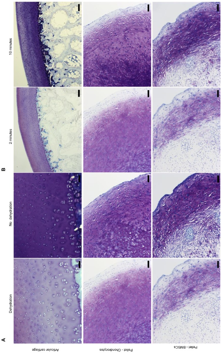
Figure. 2.
Representative images showing toluidine blue staining of sections of cartilage and of pelleted cells. (A) Panels compare effects of dehydration with no dehydration. Fixation was with 70% ethanol and sections were stained at pH 4 for 2 minutes. Scale bars 50 µm. (B) Panels compare 2 staining times. Fixation was in 70% ethanol, and sections were stained at pH 4 for 2 and 10 minutes. Scale bars: 200 µm for articular cartilage and 50 µm for pellets.

