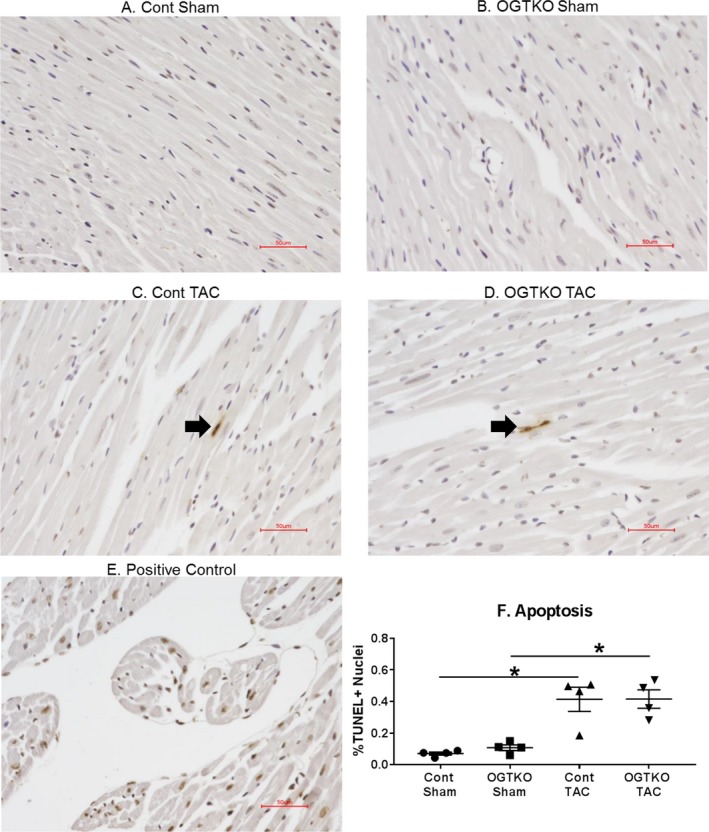
Figure 6.

O‐linked β‐N‐acetylglucosamine transferase knockout (OGTKO) does not alter histological markers of apoptosis during established hypertrophy. Sections of terminal deoxynucleotidyl transferase dUTP nick end‐labeling (TUNEL)–stained hearts from the indicated established hypertrophy groups are shown in (A through D). TUNEL‐positive nuclei in (C and D) are shown by the arrow. Because of the low number of TUNEL‐positive nuclei, these images are not representative of the overall results. E, Positive control myocardium demonstrating multiple TUNEL‐positive nuclei. F, Quantification of the percent TUNEL‐positive nuclei (% TUNEL+ nuclei) for the experimental groups. n=4 per group. Red bar indicates 50 μm. Values are expressed as mean±standard error of the mean. *P<0.05 between groups indicated with the bar. TAC indicates transverse aortic constriction.
