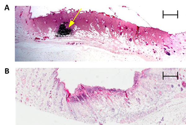
Figure 2:
Histological assessment of burn wound severity. Representative 2X hematoxylin and eosin (H&E) staining of a full thickness wound (A) and a partial thickness wound (B) 72 h post burn induction. Coagulation and red brick myocardial necrosis in the dermis and subcutaneous layers are used to classify (A) as full thickness. An intradermal fiducial marker (solid yellow arrow) is clearly visible. Scale bar, 800 μm. Revascularization of tissue as well as intact subcutaneous and muscle layers are used to confirm (B) as partial thickness. Scale bar, 600 μm.

