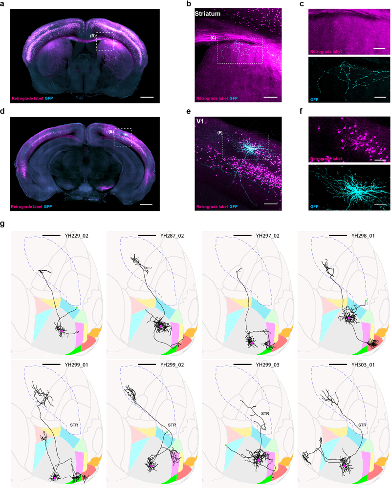
Extended Data Figure 1: Single-neuron tracing protocol efficiently fills axons projecting to the ipsilateral striatum.
We retrogradely labeled striatum projecting cells by stereotactically injecting cholera toxin subunit B conjugated with AlexaFluro594 or PRV-cre into the visual stri-atum of wild type mice or tdTomato reporter mice (Ai14, JAX), respectively (magenta). With visual guidance of two-photon microscopy, we electroporated single retrogradely labeled cells in V1 with a GFP expressing plasmid (cyan). (a) Coronal, maximum intensity projections of visual striatum. Scale bar = 1 mm. (b) Higher magnification view of the visual stratum. Scale bar = 0.2 mm. (c) Single channel images of the same axonal arbor as in (b). (d) Coronal maximum intensity projection containing V1. Scale bar = 1 mm. (e) Higher magnification view of V1. Scale bar = 0.2 mm. (f) Single channel images of V1. Scale bar = 0.2 mm. (g) Horizontal ARA-space projections of eight retrogradely labeled and electroportated cells. Cell ID numbers are indicated at the top right of each thumbnail. Scale bar = 1 mm. Note that one additional cell was retrogradely labeled and electroporated, which revealed its axonal projection to the striatum, but it is not shown because the brain was too distorted to allow accurate atlas registration.

