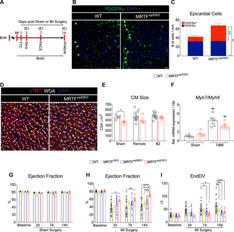
Figure 2. Preservation of cardiac physiology in MRTFepiDKO hearts after MI.
(A) Time-line analysis of adult male WT and MRTF transgenic mice subjected to Sham or MI surgery at 10–12 weeks of age. MRTF transgenic mice were analyzed by echocardiography (E) prior to surgery (baseline) and 3, 7 and 14 days after surgery. (B) Representative images of PDGFRα− and PDGFRα+ epicardial cells represented as DAPI+ nuclei outside the myocardial border (yellow arrows). (C) Quantitation of PDGFRα− and PDGFRα+ epicardial cells in 12-week old sham mice. (D) CM size was visualized in young (12-week old) sham mice. (E) Quantitation of CM crosssectional area (CSA; µm2) in sham and injured mice. (F) CM fetal gene expression of Myh7/Myh6. (G) Ejection Fraction (%) measured in male WT and MRTF transgenic mice following sham surgery. (H) Ejection fraction (%) and (I) End Diastolic Volume (µL) measurements following MI surgery.
Scale bar (B and D) = 40µm.
* p<0.05, ** p<0.01, *** p<0.001, **** p<0.0001.
## p<0.01: 7dMI versus Sham.

