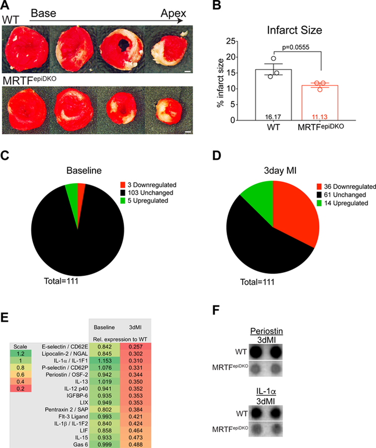
Figure 3. MRTFepiDKO mice are protected from pathological fibrotic remodeling following MI.
(A) Representative WT and MRTFepiDKO heart sections stained with TTC and quantitated in (B) 24 hours post-MI. 111 cytokines were measured and categorized as unchanged (black), upregulated (green) or downregulated (red). Screen was performed in (C) Non-injured mice and (D) Mice subjected to MI for 3-days. (E) List of downregulated cytokines in MRTFepiDKO serum relative to WT serum at baseline and 3-days post-MI. Representative dot blot images of (F) Periostin and IL-1α / IL-1F1 at 3-days post MI.
Scale bar (A) = 1mm.

