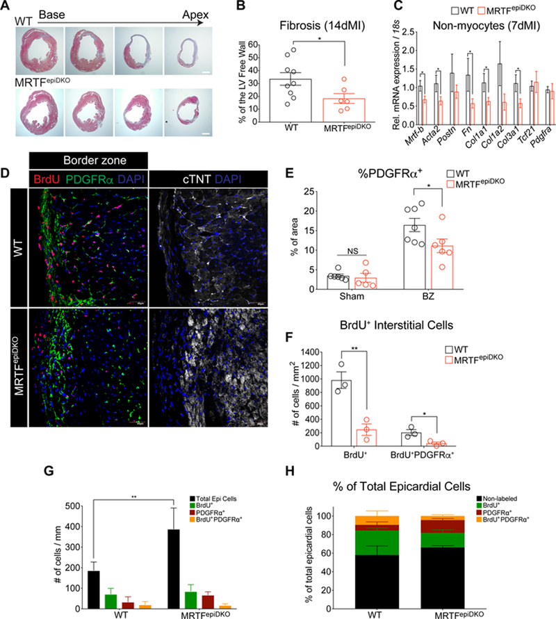
Figure 4. Cardiac fibrosis is attenuated in MRTFepiDKO hearts after MI.
(A) WT and MRTFepiDKO hearts represented in 4 layers after staining with Masson’s Trichrome. (B) Quantitation of left ventricular free wall fibrosis (%) 14 days after MI. (C) Gene expression in nonmyocytes isolated from female WT and MRTFepiDKO adult hearts 7-days post-MI. (D) Visualization of PDGFRα− and PDGFRα+ epicardial cells represented as DAPI+ nuclei outside the myocardial border and labeling of BrdU+ cells in the epicardium and interstitium in the BZ regions of the heart 14 days post-MI. (E) Quantitation of PDGFRa protein expression (% of area) located in sham and BZ regions of the heart 14 days post-injury. (F) Quantitation of the number of BrdU+ interstitial cells in the BZ regions of WT and MRTFepiDKO hearts. (G) Quantitation of epicardial cells, BrdU+, PDGFRα+., and BrdU+/PDGFRα+ epicardial cells in BZ regions of the heart. (H) Fraction of total epicardial cells (normalized to 100%) categorized as non-labeled, BrdU+, PDGFRα+, and BrdU+/PDGFRα+ in the BZ regions of the heart.
Scale bar (A) = 1mm. Scale bar (D) = 40µm.
* p<0.05, ** p<0.01.

