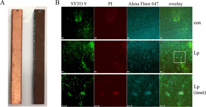
FIG 1.
Drinking water biofilms on copper (Cu) surfaces. Biofilms were established on Cu BAR slides with uncolonized and colonized slides shown on the left and right, respectively (A). Biofilms were then inoculated with LpP1s1, fluorescently stained, and visualized using confocal microscopy. (B) From left to right, membrane-intact (green, SYTO 9), membrane-permeable (red, propidium iodide [PI]), and L. pneumophila (blue, Alexa Fluor 647) cells were observed by fluorescence microscopy, including an overlay of the signals. The top row shows stained, uninoculated biofilm controls (con), the middle row shows LpP1s1-inoculated biofilms, and the bottom row shows 3×-zoomed-in area of the LpP1s1-inoculated biofilms indicated in the white box. Images are representative of four to six images collected from two independent experiments. Scale bars, 10 μm.

