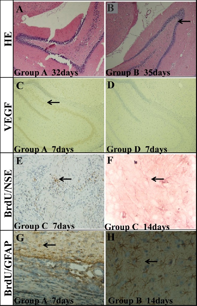
Figure 7.

Histochemical analysis of the hippocampus of TBI rats transplanted with BMSC-impregnated collagen-chitosan porous scaffold.
(A, B) HE staining in the hippocampus on the lesion side of TBI rats at 32 days (group A) (A) and 35 days (group B) (B) after BMSC transplantation. The degeneration of cells (arrow) was decreased remarkably with time. (C) Positive VEGF staining (arrow) in the hippocampus of TBI rats 7 days after BMSC transplantation (group A). (D) No VEGF staining was found in the hippocampus of group D. (E) BrdU/NSE double staining (arrow) in the damaged area 7 days after BMSC transplantation (group C). (F) BrdU/NSE double staining (arrow) in the damaged area 14 days after BMSC transplantation (group C). (G) BrdU/GFAP double staining (arrow) in the damaged area 7 days after BMSC transplantation (group A). (H) BrdU/GFAP double staining (arrow) in the damaged area 14 days after BMSC transplantation (group B). Group A: TBI + immunosuppressor + BMSCs/scaffold; group B: TBI + BMSCs/scaffold; group C: TBI + BMSCs stereotactic injection; group D: TBI only. TBI: Traumatic brain injury; BMSCs: bone marrow mesenchymal stem cells; HE: hematoxylin-eosin; VEGF: vascular endothelial growth factor; NSE: neuron specific enolase; GFAP: glial fibrillary acidic protein. Original magnifications: 100× in A–D, 40× in E and G, and 200× in F and H.
