Figure 4.
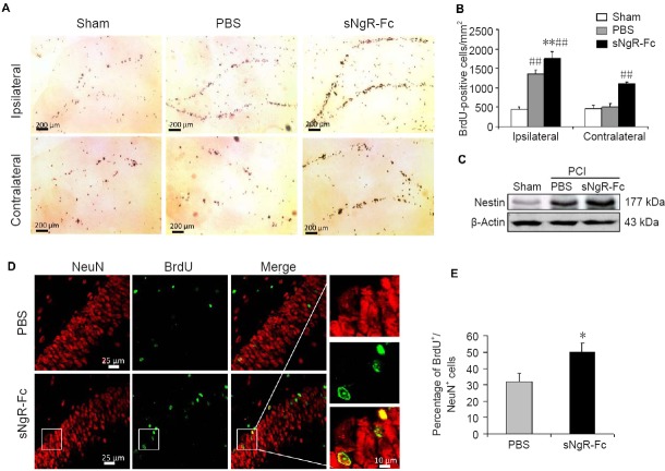
Administration of sNgR-Fc promotes the proliferation and differentiation of endogenous NPCs in rats with PCI.
(A) Representative images showing BrdU staining (brown) in the dentate gyrus on the ipsilateral (ischemic lesion) and contralateral sides after ischemic injury. Scale bars: 200 μm. (B) Quantification of BrdU-positive cells in the dentate gyrus on each side (n = 6; one-way analysis of variance followed by the Bonferroni test). (C) Nestin expression in the hippocampus measured by western blot assay. (D) Representative immunofluorescence images showing BrdU+ (green)/NeuN+ (red) cells on the side of the ischemic lesion in the PBS and sNgR-Fc groups. Scale bars: 25 and 10 μm. (E) Percentage of BrdU+/NeuN+ cells on the side of the ischemic lesion in the PBS and sNgR-Fc groups (n = 6; Student’s t-test). All data are expressed as the mean ± SD. *P < 0.05, **P < 0.001, vs. PBS group; ##P < 0.01, vs. sham group. sNgR-Fc: Soluble Nogo66 receptor-Fc protein; BrdU: 5-bromodeoxyuridine; NPCs: neural progenitor cells; PCI: photothrombotic cortical injury; PBS: phosphate-buffered saline.

