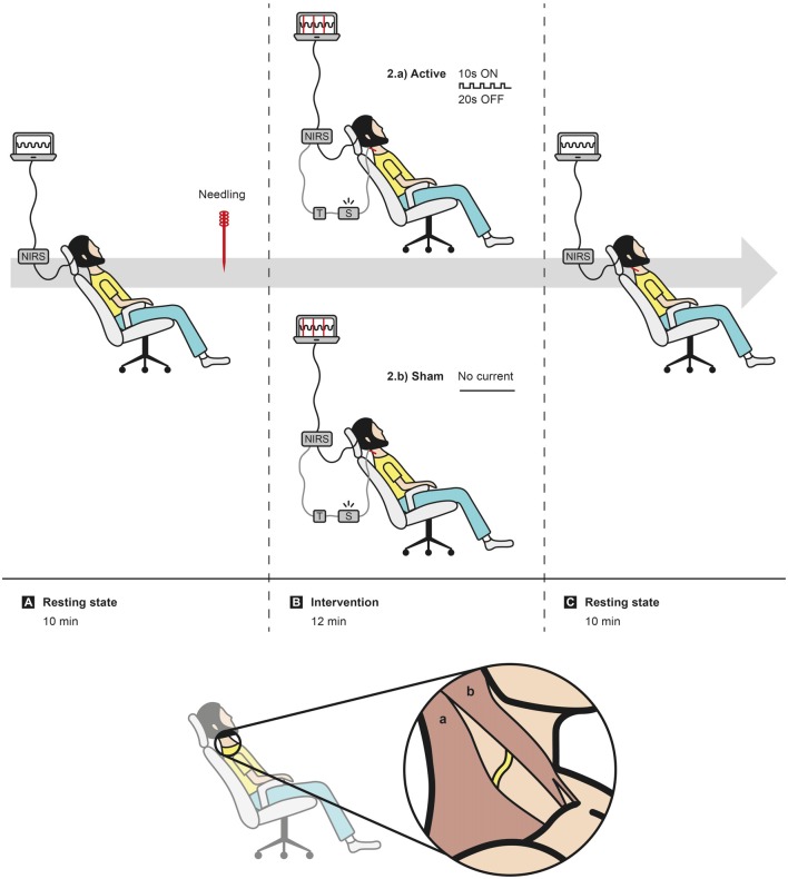
Figure 3.
Experiment layout. Sequence of events: (A) 10 min of data acquisition in resting state, followed by needling right accessory spinal nerve (ASN); (B) randomization in active or sham intervention; (C) 10 min of data acquisition in resting state. In caption, representation of the subcutaneous location of the ASN (yellow trace) between trapezius muscle (a) and sternocleidomastoid muscle (b).

