
Figure 2.
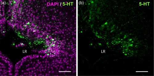
5‐HT+ CSF‐c cells in the Xenopus PVO. The laterally extended hypothalamic recess (lateral recess; LR) is visualized with DAPI staining (magenta) from a frontal section (midline to the left). CSF‐c cells immunolabeled for 5‐HT (green) are located medially in the rostral hypothalamus and laterally in the caudal hypothalamus. (a) depicts both DAPI and 5‐HT stainings, while (b) shows 5‐HT only (same picture). In the projection of confocal images (15 µm), the rostromedial and caudolateral CSF‐c cells look continuous. Scale bar = 50 µm