
Figure 9.
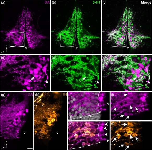
Colocalization of DA and 5‐HT in the PVO cells of zebrafish. Frontal sections of the anterior PVO (PVOa) in adult zebrafish demonstrate that CSF‐c cells are immunoreactive to DA (magenta; a, d) and to 5‐HT (green; b, e). The areas in dashed rectangles in (a), (b), and (c) are shown at higher magnification (Z‐projection = 5 µm) in (d), (e), and (f) respectively. Both monoamines were observed in a few cell bodies (arrow in f) and endfeet (arrowhead in f). In teleosts, some of the DA+ CSF‐c cells (g) are also immunoreactive for TH (h; orange). Higher magnification images of the PVO are shown in (i–l). In CSF‐c cells, intense DA immunoreactivity is present in the cell soma (j; arrows), processes, and the endfeet contacting the ventricle (k; arrowheads). In contrast, intense TH immunoreactivity is mostly observed in the soma and processes (l; arrows), but not in the endfeet. D = dorsal; L = lateral; v = ventricle. Scale bar = 50 µm in (a) (applies to a, b, c); 200 µm in (g) (applies to g, h); 100 µm in (i) (applies to i, j, k, l)