
Figure 1.
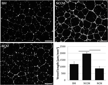
Representative images of human umbilical vein endothelial cell (HUVEC) vessel formation and vessel length measurements in base medium (BM), porcine notochordal cell‐conditioned medium (NCCM), and porcine notochordal cell‐rich matrix (NCM). Scale bars represent 250 μm. Values represent means + standard deviations, n = 5 biological repeats. Bars indicate P < 0.05 between indicated groups.