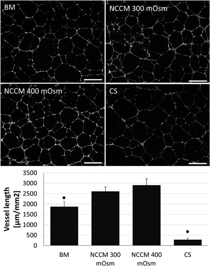
Figure 2.

Representative images of human umbilical vein endothelial cell (HUVEC) vessel formation and vessel length measurements in base medium (BM, 300 mOsm), porcine notochordal cell‐conditioned medium (NCCM) produced at approximately 300 and 400 mOsm, and with addition of an equivalent amount of chondroitin sulphate (CS). Scale bars represent 250 μm. Values represent means + standard deviations, n = 5 biological replicates. ● indicates P < 0.05 compared to all other groups.
