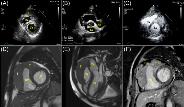
Figure 1.

A, Left ventricular short‐axis views of TTE demonstrated the tumor formation in both the RV free wall and the LV anterior wall; B, In the aortic root short‐axis view, the main pulmonary artery was narrowed; C, MCE showed rich perfusion in the thickened myocardium; D and E, The extent of the mass infiltration revealed by CMRI was consistent with TTE; F, Post‐contrast‐enhanced CMRI showed hypo‐intensive non‐homogenous gadolinium‐enhanced masses, focal areas of non‐enhancement (asterisk) implied pericardial effusion. (T indicates tumor; LV, left ventricle; RV, right ventricle; AO, aortic; LA, left atrial)
