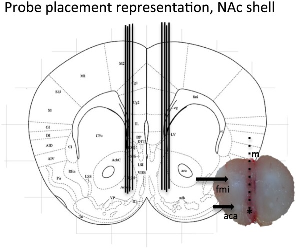
Figure 1.

Representative brain slice and schematic representation of probe placement in nucleus accumbens (NAc) shell. A coronal mouse brain section showing eight representative probe placements (illustrated by vertical lines) in the NAc shell. One brain slice shows a representative probe placement in the NAc shell. The asterisk shows the targeted area; vertical lines represent the probe; m, midline; fmi, anterior forceps of corpus callosum; aca, anterior commissure
