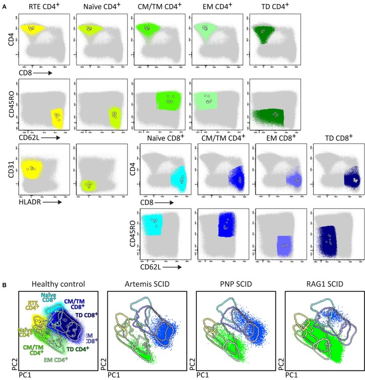
Figure 6.
Application of the RTE-SCID tube in healthy controls and T-cell defects. Analysis of circulating T-cell subsets (FS/SSloCD3+) identified within 1x10⁶ blood leukocytes using the markers from the RTE-SCID tube: Recent Thymic Emigrants (RTE) CD4+ T-cells (CD4+ CD8− CD45RO− CD62L+ CD31+ HLA-DR−), and naïve (CD45RO− CD62L+ CD31+ HLA-DR−), Central/Transitional Memory (CM/TM) (CD45RO+ CD62L+), Effector Memory (EM) (CD45RO+ CD62L−), and Terminally Differentiated (TD) (CD45RO− CD62L−) CD4+ and CD8+ T-cells. (A) The identification of biologically relevant T-cell subsets in six healthy controls using the minimum number of bivariate plots required for a 6 marker-combination (three bivariate plots/cell population). (B) Comparable T-cell data via PCA1 vs. PCA2 representation of a 6-dimensional space in six healthy controls (left) as well as in three severe combined immunodeficiency (SCID) patients, diagnosed with Artemis, PNP and RAG-1 defects, all stained with the RTE/SCID tube under comparable conditions. All three SCID patients show “leakiness” with virtually complete absence of naïve T-cells (<1 cell/μL blood) and clear “right shift” to mature CM/TM, EM, and TD T-cells in both the CD4 and CD8 lineages.

