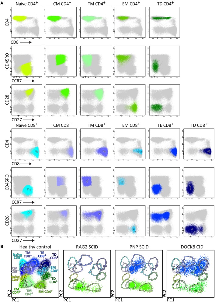
Figure 7.
Application of the T-cell panel tube in healthy controls and PID patients with T-cell defects. Analysis of circulating T-cell subsets (FS/SSloCD3+) identified within 1 × 10⁶ blood leukocytes using the markers from the T-cell panel tube: naïve (CD45RO− CCR7+ CD27+ CD28+), Central Memory (CM) (CD45RO+ CCR7+ CD27+ CD28+), Transitional Memory (TM) (CD45RO+ CCR7− CD27+ CD28+/−), Effector Memory (EM) (CD45RO+ CCR7− CD27− CD28+), Terminal Effector (TE) (CD45RO− CCR7− CD27+ CD28+/−), and Terminally Differentiated (TD) (CD45RO− CCR7− CD27− CD28+/−) CD4+ and CD8+ T-cells. (A) The identification of biologically relevant subsets of T-cells in six healthy controls using the minimum number of bivariate plots required for a 6 marker-combination (three bivariate plots/cell population). (B) Comparable data via PCA1 vs. PCA2 representation of a 6-dimensional space in blood of six healthy controls (left) as compared to blood samples of three severe combined immunodeficiency (SCID) patients, diagnosed with RAG2, PNP, and DOCK8 defects, all stained with the RTE/SICD tube under comparable conditions. The SCID patients show “leakiness” with absence of naïve T-cells (<1 cell/μL blood) and variable “right shift” to mature CM/TM, EM, and TD T-cells in the CD4 lineage (all 3 SCID patients) and CD8 lineage (PNP and DOCK8 defects).

