
Figure 1.
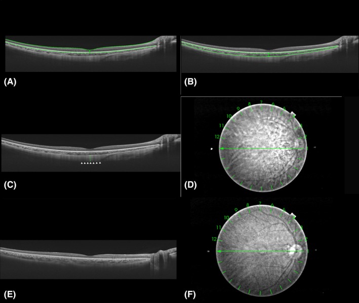
Cross‐sectional images collected by swept‐source optical coherence tomography and thickness measurements of each layer at the centre of the fovea. Horizontal cross‐sectional images of the posterior pole of the same participant were collected by swept‐source optical coherence tomography (SS‐OCT). Subfoveal retinal, choroidal and scleral thicknesses were manually measured at the centre of the fovea. (A) The inner border (internal limiting membrane, indicated by the upper horizontal green line) and outer border [the outer border of the retinal pigment epithelium (RPE), indicated by the lower horizontal green line] of the retina. Retinal thickness was defined as the vertical distance between the two borders (indicated by the vertical green line). (B) The inner border (the outer border of the RPE, indicated by the upper horizontal green line) and outer border (choroidal–scleral interface, indicated by the lower horizontal green line) of the choroid. Choroidal thickness was defined as the vertical distance between the two borders (indicated by the vertical green line). (C) The outer border of sclera (indicated by the white arrowheads). Scleral thickness was defined as the vertical distance between the choroidal–scleral interface and the outer scleral border (indicated by the vertical green line). (D) The fundus of A, B and C. Measurements were performed on the vertical and horizontal lines (line 1 and line 7). (E) Image with unclear scleral border. (F) The fundus of E.