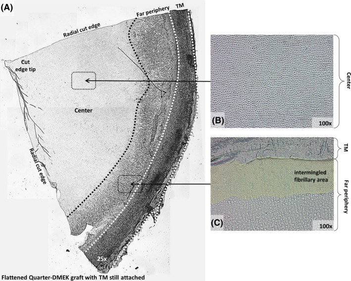
Figure 1.

General view of a Quarter‐Descemet membrane endothelial keratoplasty (DMEK) graft flattened on a glass support. (A) Overview of a flattened Quarter‐DMEK graft with the trabecular meshwork (TM) still attached. Corneal centre (B) and far periphery (C) show structural differences, which become more distinct when displayed at higher magnifications (×100). (B) Corneal centre with closely packed hexagonal endothelial cells. (C) Far peripheral area is dominated by a fibrillary area adjacent to the TM.
