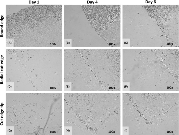
Figure 3.

Example of collective in vitro endothelial cell (EC) migration. (A–I) Light microscopy images of the round edge (A–C), the radial cut edge (D–F) and the cut edge tip (G–I) of the Quarter‐Descemet membrane endothelial keratoplasty (DMEK) graft taken at Day 1 (left), Day 4 (middle) and Day 6 (right) with ×100 magnification. (A–C) In the area of the round edge and far periphery of the Quarter‐DMEK graft, no EC migration onto the glass slide was observed up to Day 6. (D–F) Along the radial cut edges of the Quarter‐DMEK graft, collective EC migration in a form of a monolayer was observed; leader cells at the front edge of the advancing cell sheet are identifiable. (G–I) Around the cut edge tip of the Quarter‐DMEK graft, the collective migration pattern was most evident at Day 6.
