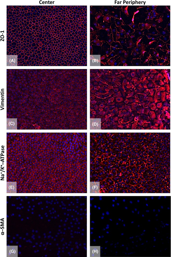
Figure 4.

Immunofluorescence staining of the Quarter‐Descemet membrane endothelial keratoplasty (DMEK) graft in the centre compared to the far periphery. Expression of ZO‐1 (A,B), vimentin (C,D), Na+/K+‐ATPase (E,F) and α‐smooth muscle actin (α‐SMA; G,H) was analysed. The central endothelium showed characteristic expressions for the tight junction protein ZO‐1 (A, red), structural protein vimentin (C, red) and functional protein Na+/K+‐ATPase (E, red) counterstained with 4′,6‐diamidino‐2‐phenylindole (blue). The presence of markers in the far peripheral area (B,D,F) verified the presence of endothelial cells (ECs) up to the round edge of the Quarter‐DMEK graft. However, the cells in the far periphery showed a different expression pattern for these endothelial markers (B,D,F red) as compared to the central area. α‐SMA, used as a negative control for the ECs, was absent in the centre and in the far periphery of the endothelium (G,H). ×200 magnification.
