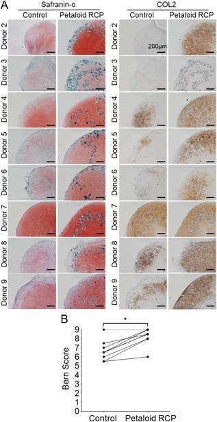
Figure 4.

Histological appearance of the cartilage pellet derived from synovial MSCs cultured with or without petaloid RCP. An initial 1.25 × 10⁵ synovial MSCs derived from donors 2 to 9 were cultured with or without petaloid RCP in chondrogenic medium for 21 days. (A) Histological appearance of the cartilage pellets stained with safranin‐o and immunostained for type II collagen. Bars = 200µm. (B) Bern score (0–9) for evaluation of the safranin‐o stained cartilage pellet. *p < 0.05 by Wilcoxon matched‐pairs signed‐rank test.
