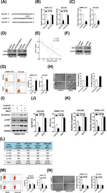
Figure 5.

RAC1 was a direct target of miR‐365 in liver CSCs. (A) A potential target site for miR‐365 in the 3′‐UTR of human RAC1 mRNA, as predicted by the program Targetscan. To disrupt the interaction between miR‐365 and RAC1 mRNA, the target site was mutated. (B) Luciferase reporter assays performed in SMMC7721 miR‐365 or HCCLM3 miR‐365 and their control cells transfected with wild‐type or mutant RAC1 3′‐UTR constructs. (C) The mRNA expression of RAC1 was checked in SMMC7721 miR‐365 or HCCLM3 miR‐365 and their control cells by real‐time PCR. (D) The protein expression of RAC1 was checked in miR‐365 overexpression or miR‐365 inhibitor and control cells by Western blot. (E) Spearman correlation analysis of the relationship between RAC1 protein and miR‐365 expression in 30 HCC specimens. (F) HCCLM3 and SMMC7721 cells were infected with RAC1 overexpression virus and the sable infectants were checked by Western bolt assay. (G) Flow cytometric analysis of the proportion of CD133+ or EpCAM+ cells in RAC1 overexpression and control cells. (H) Spheres formation assay of RAC1 overexpression and control hepatoma cells. (I) The expression of p‐ERK, p‐AKT and β‐catenin was checked in miR‐365/RAC1 overexpression HCC cells. (J) HCCLM3 miR‐365 or SMMC7721 miR‐365 and their control cells were infected with RAC1 overexpression virus or control virus and the EpCAM+ or CD133+ hepatoma cells was checked by flow‐cytometric assay. (K) SMMC7721 miR‐365 or HCCLM3 miR‐365 and their control cells were infected with RAC1 overexpression virus or control virus and subjected to spheroid formation. (L) In vivo limiting dilution assay of indicated HCC cells. Tumors were observed over 2 months; n = 8 for each group. (M) HCCLM3 miR‐365 or SMMC7721 miR‐365 and their control cells were treated with EHop‐016 (10 µM) or not and the EpCAM+ or CD133+ hepatoma cells was checked by flow‐cytometric assay. (N) SMMC7721 miR‐365 or HCCLM3 miR‐365 and their control cells were treated with EHop‐016 (10 µM) or not and subjected to spheroid formation
