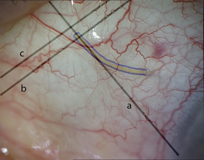
Figure 1.

Location of anterior segment optical coherence tomography (AS‐OCT) scans for assessment of bleb morphology after XEN Glaucoma Gel Microstent (XEN‐GGM) implantation. Slit lamp photograph of a bleb 1 week after XEN‐GGM implantation. The XEN‐GGM is highlighted in blue. One scan was obtained radially to the limbus and through the outer part of the XEN‐GGM (a). A second scan perpendicular to the first one was taken to image the subconjunctival part of the XEN‐GGM close to its exit site (b). A third scan was taken parallel and 1 mm posterior to the outer XEN‐GGM lumen through the site of maximal bleb elevation (c).
