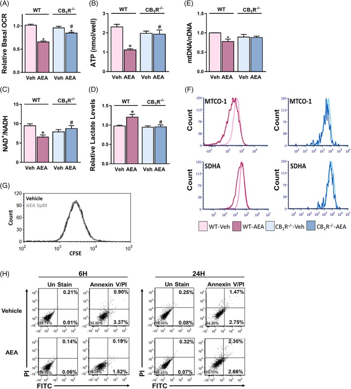
Figure 2.

Acute activation of CB1R in HK‐2 cells impairs mitochondrial function and biogenesis. WT‐HK‐2 or CB1R‐/‐‐HK‐2 cells were treated with either vehicle (Veh) or 5 μM AEA for 6 hours and were then analysed for mitochondrial function. A, Reduced oxygen consumption rate (OCR) measured using the Seahorse XF analyzer, B, reduced ATP levels, and C, reduced NAD+/NADH ratio, as well as D, elevated cellular lactate levels, were found in WT‐HK‐2 cells treated with AEA (5 μM). Data represent the mean ± SEM of at least six replicates from three independent experiments. E, The ratio between mitochondrial and nuclear DNA. F, FACS analysis for MTCO‐1 and SDHA, demonstrating reduced biogenesis following treatment of wild‐type cells with AEA. G, H, WT‐HK‐2 cells, exposed to Veh or 5 μM AEA, were analysed by flow cytometry for proliferation (G) with CFSE staining after 24 hours of treatment or for apoptosis (H) with Annexin V and Propidium Iodide at the indicated time points. Data represent one experiment out of three performed (in triplicate). *P < 0.05 relative to Veh‐treated HK‐2 of the same cell line. # P < 0.05 relative to the same treated group in WT‐HK‐2 cells
