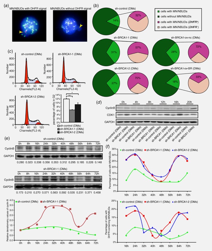
Figure 4.

HR inhibition results in G2/M abrogation and cell cycle acceleration accompanied by promoting the exclusion of DMs via MN/NBUDs. a. FISH analyses of MN/NBUDs in DM‐containing control and BRCA1‐depleted cells probed with BAC‐containing DHFR and the centromere of chromosome 5. The yellow arrow indicated the MN/NBUDs of nuclei. The MN/NBUDs were grouped into two categories: with DHFR signal (left panel) and without DHFR signal (right panel; DHFR in green; centromere of chromosome 5 in red; DAPI in blue). b. analyses of MN/NBUDs formation and exclusion of DHFR via MN/NBUDs in DM‐containing control, two BRCA1‐depleted clones, BRCA1‐depleted control and BRCA1‐depleted rescued clone. (**p < 0.005 for MN/NBUDs formation between control and two BRCA1‐depleted clones, n ≥ 100; **p < 0.005 for MN/NBUDs formation between BRCA1‐depleted control and BRCA1‐depleted rescued clone, n ≥ 100; *p < 0.025 for exclusion of DHFR via MN/NBUDs between control and two BRCA1‐depleted clones; *p < 0.025 for exclusion of DHFR via MN/NBUDs between BRCA1‐depleted control and BRCA1‐depleted rescued clone.) c. Flow assay analyses of cell cycle distribution in DM‐containing control and two BRCA1‐depleted clones. The left panel shows distributions of G1, S and G2 phases. The right panel shows both the G2 phase percentage and cell number of G2 phase for 3 repetitions. (*p < 0.025, **p < 0.005, by Chi‐squared test and Bonferroni adjustment) d. Western blot of cyclin B and CDK1 in DM‐containing control and BRCA1‐depleted cells harvested at different time points of releasing in complete culture (0 to 20 h) and then recorded once every 4 h. E. Western blot analyses of cyclin B in DM‐containing control and BRCA1‐depleted cells released at different time points of releasing in complete culture (0 to 72 h) and recorded once every 8 h. The numbers under the bands represent the relative densitometry values. The lower panel showed the trends of cyclin B expression in DM‐containing control and BRCA1‐depleted cells. f. Analyses of MN/NBUDs formation and exclusion of DHFR via MN/NBUDs in DM‐containing control and two BRCA1‐depleted clones harvested at different time points of releasing in complete culture (16 to 72 hr) and recorded once every 8 h.
