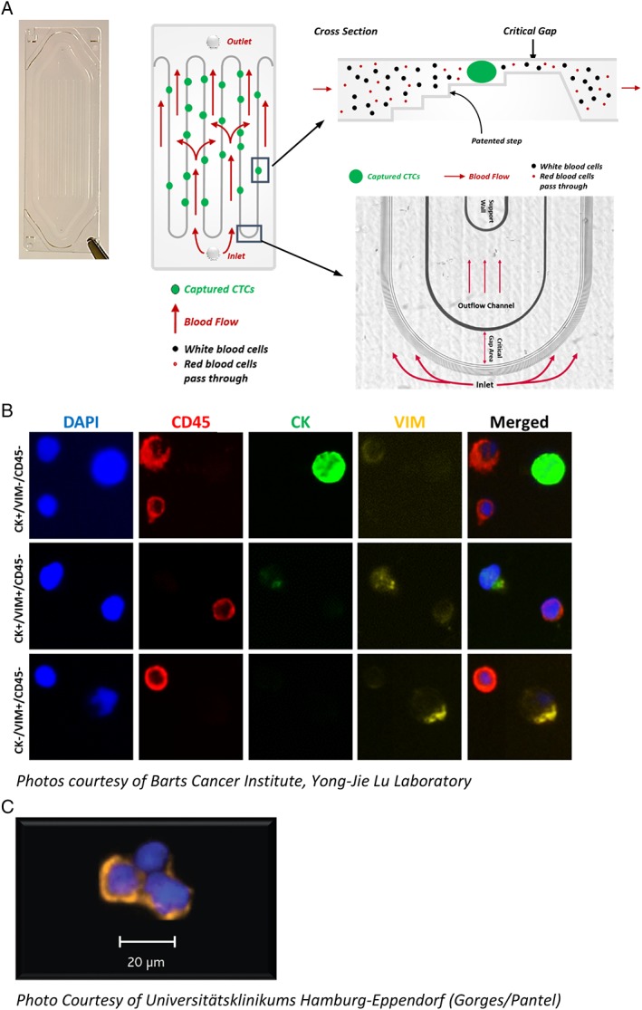
Figure 2.

(A) Image and diagram of a Parsortix GEN3 Cell Separation Cassette showing how the blood flows into the cassette, over the step structures and through the critical gap. (B) Example images of nucleated blood cells and CTCs harvested from prostate cancer patients (7.5 ml EDTA blood samples processed within 4 h after collection) and immunofluorescently stained with DAPI (blue), CD45 (red), cytokeratin (green), and vimentin (yellow). (C) Example image of CTCs harvested from breast cancer patient (4 ml CellSave blood sample processed <24 h after collection) and immunofluorescently stained with cytokeratin (orange) and DAPI (blue).
