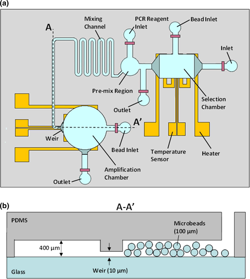
Fig. 2.

Schematics of the aptamer isolation microchip. a Top view showing microchambers for affinity selection and PCR amplification. These chambers are connected via a serpentine diffusion-based mixing channel and integrated with thin-film resistive heaters and temperature sensors. b Cross-sectional view showing a weir-like restriction in which openings of 10 μm height retain microbeads roughly 100 μm in diameter
