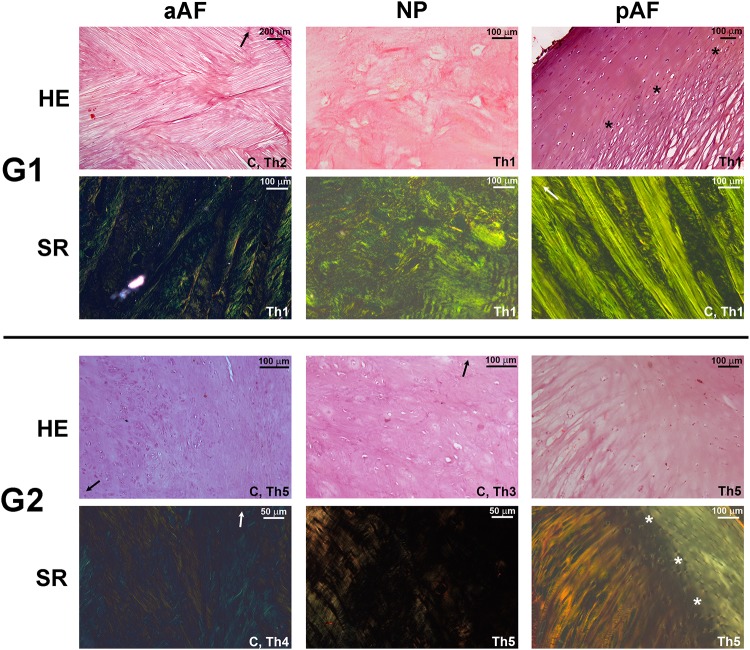
Fig 3. Light microscopy images.
Alternating diagonal and longitudinal lamellae are visible in the aAF and predominantly longitudinal in the pAF. Sharpey-type insertion of these fibers into the endplate is visualized in G1 (asterisks) and G2. The loose fibrocartilaginous phenotype of the G1 AF is substituted for a cartilaginous one in G2 with dense extracellular matrix and chondrocyte clusters. HE, hematoxylin & eosin. SR, Sirius Red. “C” designates a coronally-oriented section, Th notes Thompson grade, arrow indicates the nearest endplate.

