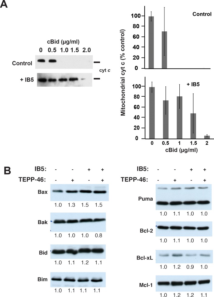
Fig 6. Mitochondria from cells expressing IB5 were relatively resistant to cBid-induced MOMP.
A. Cyt c release assay. Left panel: control (top) or intrabody-expressing (bottom) 293T cells were collected, and the mitochondrial fraction was isolated by differential centrifugation. To induce MOMP, recombinant cBid protein was added at the indicated concentrations. After incubation for 30 min at 37 ºC, samples were centrifuged, and cyt c content in mitochondrial pellet fractions was analyzed by immunoblot. A representative of three independent experiments is shown. Right panel: densitometric quantification of average cyt c content ± SEM from three independent experiments. B. Levels of several Bcl-2 family proteins were unchanged following IB5 expression or incubation with TEPP-46 or both. Cell lysates from 293T cells infected with and without IB5 and incubated with and without TEPP-46 (27 μM) were separated on SDS-12% polyacrylamide gels. Bcl-2 family proteins were detected by immunoblotting. The bands were quantified using ImageJ and normalized to the control cell lysate on the leftmost lane. Note: underlying data are included in corresponding tabs in the accompanying supplemental Excel file, S1 Data. 293T, HEK293T; cBid, cleaved Bid; cyt c, cytochrome c; IB5, intrabody 5; MOMP, mitochondrial outer membrane permeabilization.

