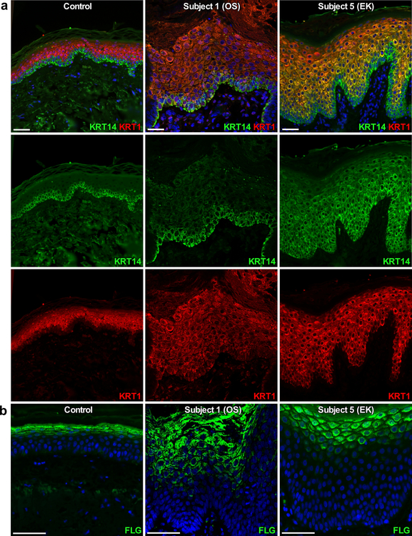
Figure 4. Immunostaining of skin from subjects with PERP mutations for markers of epidermal differentiation.
DAPI nuclear counterstain is in blue; scale bars are 50 μm. Left panels are control tissue (age 32, abdominal biopsy), middle panels are OS Subject 1 (p.Tyr153* heterozygote), and right panels are EK Subject 5 (p.Ser38Leufs*52 homozygote). (a) Positive immunostaining for keratin 14 (KRT14, green) is limited to the epidermal basal layer in the control and Subject 1 but is expanded in Subject 5, while positive immunostaining for keratin 1 (KRT1, red) is limited to suprabasal epidermis in both control and Subjects 1 and 5. (b) Immunostaining for filaggrin (FLG, green) is tightly restricted to the epidermal granular layer in the control, but is expanded in Subjects 1 and 5.

