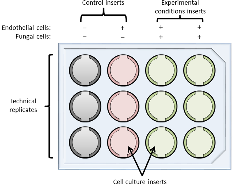
Figure 1: Layout for a typical experiment testing two experimental conditions with experimental and control wells in triplicate.

1st column, empty insert controls used to measure background values for TEER calculations. 2nd column, inserts with endothelial cells alone, to assess baseline TEER (in the absence of fungi) that can be used to correct all other TEER values. 3rd and 4th columns, inserts with both endothelial and fungal cells. The last two columns may be used to assess any alteration in TEER induced by the fungi (by comparing TEER values to those for the 2nd column) and also to measure fungal traversal under the desired experimental conditions, for example opsonized (column 3) versus non-opsonized fungal cells (column 4).
