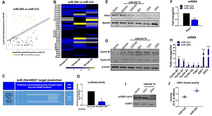
Figure 4: Analysis of molecular signaling in miR-294 treated NRVMs.
A) Cell cycle array analysis of NRVMs with miR-294–3p mimic or control treatment from 3 independent experiments (n=3). B) Heat map representation of miR-294 fold change over miR-Ctrl. C) Target prediction analysis for miR-294 putative sites on 3-UTR of target genes, identified a 7mer-8m target site in 3-UTR of Wee1 (position 493–499). D) Dual luciferase reporter activity assay for validation of miR-294 targeting of 3UTR-Wee1 in the presence of miR-294 mimic indicates reduced luciferase activity in NRVMs transfected with plasmid carrying 3-UTR-Wee1 (n=3). E) Immunoblot analysis show reduced Wee1 expression in NRVMs after treatment with miR-294 mimic together with mRNA expression in (F) (n=3). G) Elevated proteins levels for cyclin B1 and D1 in miR-294 treated NRVMs (n=3). H) miR-294 treatment of NRVMs shows increased mRNA expression of Cyclin B1,D1, E1, A2, CDK1, E2F1 and E2F3 compared to control cells (n=3). I) Decreased phosphorylation of CDK1tyr15 to total CDK1 ratio (n=3). (J) Elevated CDK1 kinase activity in NRVMs treated with miR-294 compared to control (n=3). miR-Ctrl vs. miR-294 *p < 0.05, **p < 0.01, ***p < 0.001, data was assessed using unpaired student’s t test.

