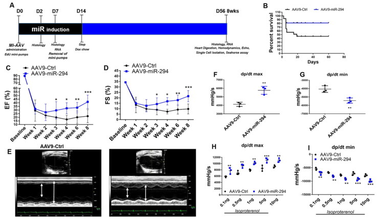
Figure 5: AAV9-miR-294 administration augments cardiac function after myocardial infarction in mice.
A) Schematic illustration of experimental design. Animals were divided into two groups and administered AAV9-GFP (n=20) and AAV9-miR-294 (n=20) two weeks prior to myocardial infarction surgery. At the day of surgery (D0) mice were subjected to LAD ligation followed by administration of doxycycline chow to induce miR-294 expression that was removed at day14 and EdU mini-pump implantation removed at D7. Hearts were harvested at D2 and D7 for histology and RNA analysis. Heart were analyzed for echos/hemodynamic measurements, histology and RNA analysis at terminal time point was 8 weeks or D56 after MI. B) Kaplan-Meir survival curve analysis shows increased %survival in AAV9-miR-294 group compared to AAV9-GFP. C) Increased ejection fraction and fractional shortening (D) in AAV9-miR-294 (n=10) administered hearts compared to AAV9-GFP (n=8) animals at 8 weeks after MI. E) Increased wall contractility in AAV9-miR-294 animals compared to controls. Hemodynamic measurements show increased dp/dtmax and reduced dp/dtmin under baseline (F-G) and stimulated (H-I) conditions 8 weeks after MI (n=3). AAV9-Ctrl vs. AAV9-miR-294 *p < 0.05, **p < 0.01, ***p < 0.001, data in panels C,D, were assessed using one-way analysis of variance H and I was assessed using two-way ANOVA with bonferroni post-hoc test while panels F and G were assessed using unpaired student’s t test.

