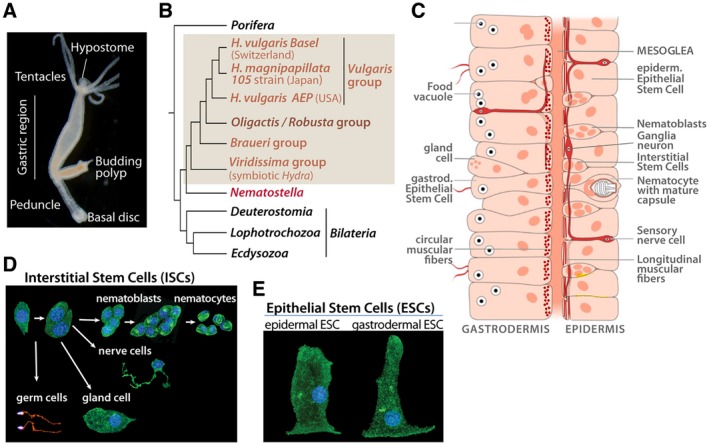
Figure 1.

Hydra anatomy, phylogenetic position and cell types. (A) Bright field picture of a budding H. oligactis. Note the typical stalk shape of the peduncle region. (B) Phylogenetic tree showing the respective positions of H. vulgaris and H. oligactis. (C) Schematic view of the bilayered organization of the body column with the gastrodermis (or gut) on the left side and the epidermis on the right one, separated by an acellular matrix named mesoglea. Epithelial cells from both layers contain myofibrils, circular in gESCs and longitudinal in eESCs. Nematoblasts and nematocytes are restricted to the epidermis while nerve cells are present in both layers although more abundant in the outer one. Scheme modified from (Lenhoff and Lenhoff, 1988). (D, E) Identification of the various Hydra cell types, as observed after tissue maceration followed by b‐tubulin immunodetection and DAPI counter staining. Among the derivatives of the multipotent ISCs (D), note the precursors to nematocytes (or cnidocytes) named nematoblasts that divide syncitially before entering differentiation, and the different types of nerve cells: sensory, sensory/motor (bipolar), or ganglion neurons (multipolar). The ESCs (E) are unusual stem cells, that is, self‐renewing and differentiated (Buzgariu et al., 2015). [Colour figure can be viewed at wileyonlinelibrary.com]
