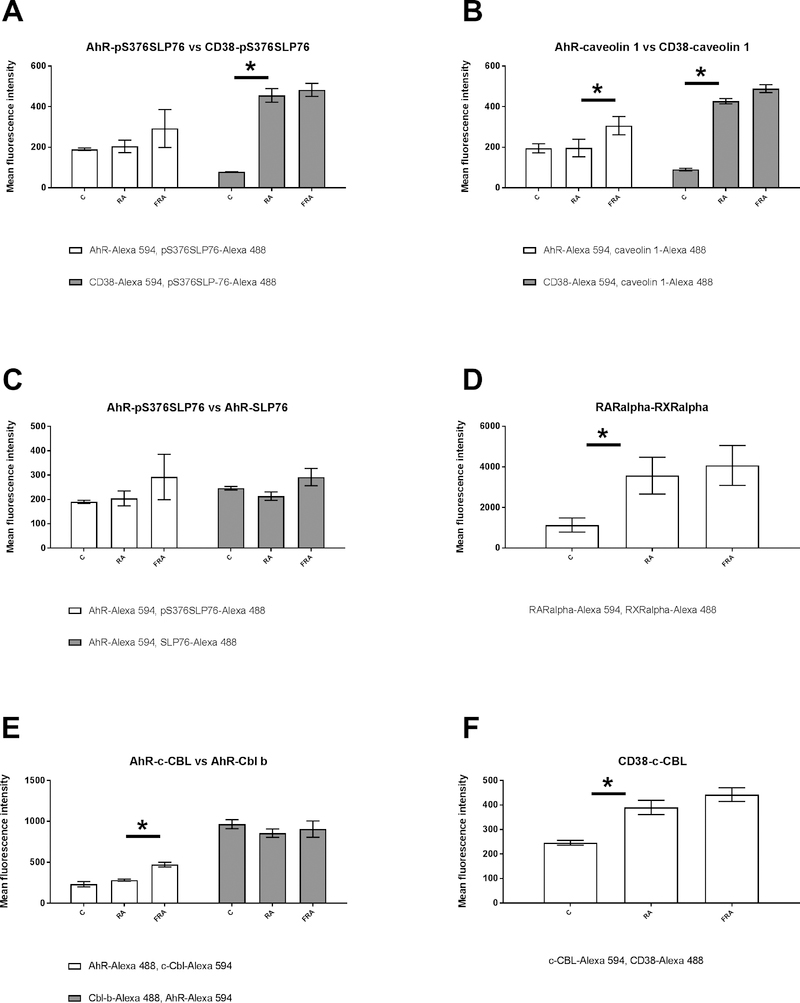
Figure 1: RA and RA+FICZ modulation of protein-protein associations in the RA induced signalsome.
HL-60 cells were initiated in culture at 0.1 × 10⁶ cells/ml and treated with 1 μM RA, 100 nM FICZ, as indicated. Cells were harvested, fixed for 10 min with 2% paraformaldehyde, and permeabilized with ice cold methanol. Cells were labeled with primary antibodies (or isotype controls) directly conjugated to Alexa Fluor 488 or 594, as indicated. The immunocomplexes were analyzed using flow cytometry (BD FACS Aria III SORP, BD Biosciences). Mean fluorescence intensity for the FRET signal is presented. (A) comparison of AhR-pS376SLP76 and CD38-pS376SLP76 FRET for control, RA and RA+FICZ treated cells. (B) comparison of caveolin-1 – AhR and caveolin-1- CD38 interactions. (C) comparison of AhR interactions with either SLP76 or pS376SLP76. (D) comparison of RARα-RXRα interactions. (E) comparison of AhR interactions with either c-Cbl or Cbl b.(F) comparison of CD38 interactions with c-Cbl.

