Figure 1.
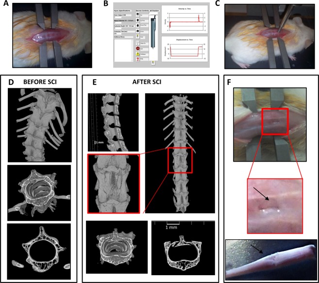
Generation of severe spinal cord injury. (A) Representative example of the exposition of the spinal cord in anesthetized animal mounted on a stereotaxic apparatus. (B) Spinal adaptors are connected to a cortical PinPoint precision impactor device (Stoelting), being the impactor positioned on T9-T11 vertebrae. The graphical impact parameters are used to identify potential outliers. To obtain a severe spinal trauma the following parameters are set: - middle, round and flat tip (#4); - velocity 3 m/sec; - depth 5 mm; - dwell time 800 ms. (C) Representative example of the impactor on the spinal cord of an anesthetized animal. (D) Representative longitudinal and cross-sectional μCT images of thoracic spinal cord in intact and (E) in SCI mice, magnification of the impact zone at the thoracic level after SCI is shown in the square. (F) Impact generates a perforation of vertebrae bones and some fragments are visible (zoom, arrow). After impact spinal cord appears compressed (bottom).

