Figure 3.
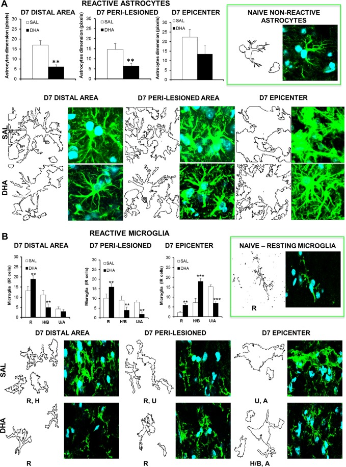
Morphometric analysis of astrocytes and microglia after SCI. Representative high magnification (63X, zoom 3X) images of reactive astrocytes (A) and microglia (B) at distal, peri-lesioned and epicenter areas of spinal injured tissues, in saline (SAL) and DHA-treated mice, 7 days (D7) after SCI (n = 3 mice/group). Each image was transformed in digital image, where outline of cell silhouettes were identified and automatically measured for astrocytes, while microglia were singularly counted and divided about the different morphology. (A) Note the increase of GFAP expression and morphological changes showing hypertrophic status of astrocytes, with glial scar formation in the epicenter area, in comparison with naïve non-reactive astrocytes (in the green frame). As shown in graphs, DHA significantly reduced the dimension of astrocytes (quantified by using RGB method that converted pixel in brightness values) in distal and peri-lesioned areas and, even if not statistically significant, in the epicenter, where also phenotypic changes occurred in comparison with SAL-injected mice. (B) After SCI microglia show a hyperactive condition characterized by several phenotypes, differently distributed in the analysed areas: from resting (R) to hypertrophic/bushy (H/B) and un-ramified/amoeboid (U/A) states. As evidenced in graphs, DHA significantly increased the R state in all areas investigated. The increase of the H/B phenotype observed at the epicenter supports phagocytic activity, which may contribute to homeostasis reinstatement. **p < 0.01, ***p < 0.001 vs SAL Student’s t test.

