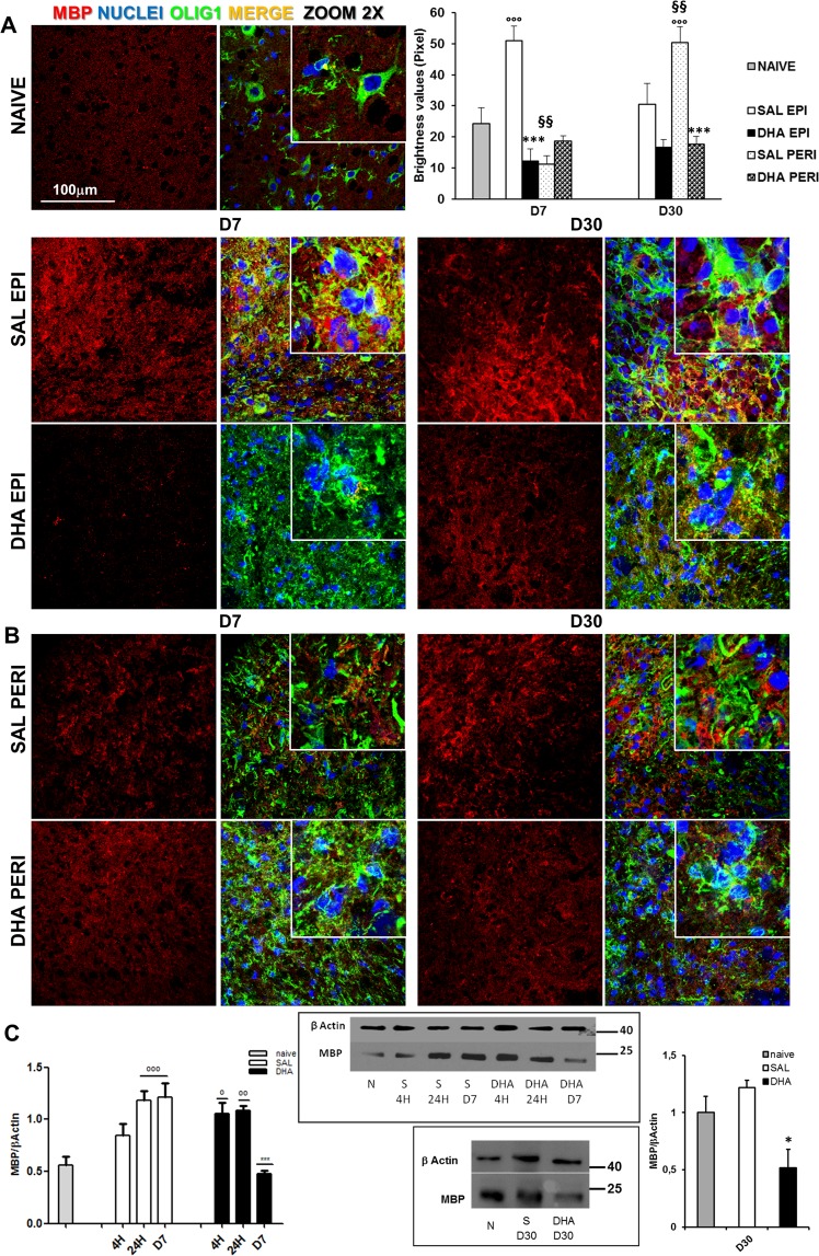
Figure 4.
Myelination after SCI: Expression of Myelin Basic Protein. (A) Representative images (63X, zoom 2) of MBP expression in naïve and saline (SAL)- and DHA-treated mice at the epicenter (EPI) and (B) peri-lesioned (PERI) areas, 7 (D7) and 30 days (D30) after SCI. The quantification of MBP, in graph, showed significant increase of MBP expression in SAL EPI at D7 and SAL PERI at D30 in comparison with naïve, while DHA significantly differed from SAL (n = 3 mice/group) (°°°p < 0.001 vs naïve; ***p < 0.001 vs SAL; §§p < 0.01 SAL PERI vs SAL EPI. Tukey/Kramer). (C) Western blot analysis and quantification of MBP in naïve/N, saline (S)(SAL) and DHA-treated mice 4 hours (4 H), 24 hours (24 H) and 7 days (D7) after SCI. The time dependent increase of MBP in injured tissues of SAL mice was gradually reduced in DHA-treated animals, where the expression of MBP reached the naïve values at D7. At D30 (graph on the right) MBP expression in DHA is significantly reduced compared to SAL-treated samples (n = 3 mice/group/time point). (°p < 0.05, °°p < 0.01, °°°p < 0.001 vs naïve; *p < 0.05, ***p < 0.001 vs SAL; Dunn’s Test).

