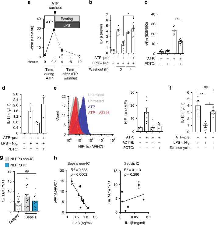
Fig. 7.
Mitochondrial dysfunction mediates P2X7 receptor-induced NLRP3 inflammasome impairment. a Mitochondrial membrane depolarization in BMDMs treated with ATP (3 mM, 30 min), then washed-out and incubated for the indicated times with or without LPS (1 μg/ml). b IL-1β release from wild-type BMDMs treated as in a, but after LPS priming cells were stimulated with nigericin (10 μM, 30 min). c Mitochondrial membrane depolarization in BMDMs treated with ATP (3 mM, 30 min) in the presence or absence pyrrolidine dithiocarbamate (PDTC, 40 μM). d IL-1β release from PBMC isolated from healthy donor blood samples treated with ATP (1 mM, 30 min; ATP-pre) with or without PDTC (10 μM), then washed and primed with or without LPS (1 μg/ml, 4 h) and then stimulated with nigericin (10 μM, 30 min). e Representative histogram plot of HIF-1α staining (left) or quantification of mean intensity fluorescence increase (ΔMFI, right) in monocytes from healthy donor blood samples treated or not with ATP (1 mM, 30 min) in the presence or absence of AZ11645373 (10 μM) or PDTC (10 μM), and then washed and cultured for 2 h; non-stained monocytes (light gray, left). f IL-1β release from BMDM supernatants treated with ATP (3 mM, 30 min; ATP-pre) in the presence or absence of echinomycin (5 nM), then washed and primed with or without LPS (1 μg/ml, 4 h) and then stimulated with nigericin (10 μM, 30 min). g Expression of HIF1A analyzed by qPCR from control surgery group and septic patients PBMCs; septic patients are separated into NLRP3 non-immunocompromised (gray bar) and immunocompromised (blue bar). h Correlation between HIF1A expression and IL-1β released from septic patients PBMCs treated with LPS (1 μg/ml, 2 h) and ATP (3 mM, 30 min); IC: immunocompromised septic patients. Each dot represents a single independent experiment or a sample from an individual healthy donor or septic patient; average ± standard error is represented in panels a–g; exact n number for each panel is presented in Source Data file; *p < 0.05; **p < 0.01; ***p < 0.001; ns, no significant difference (p > 0.05); Kruskal–Wallis test was used for b, c, f; Pearson correlation was used in h

