Figure 5.
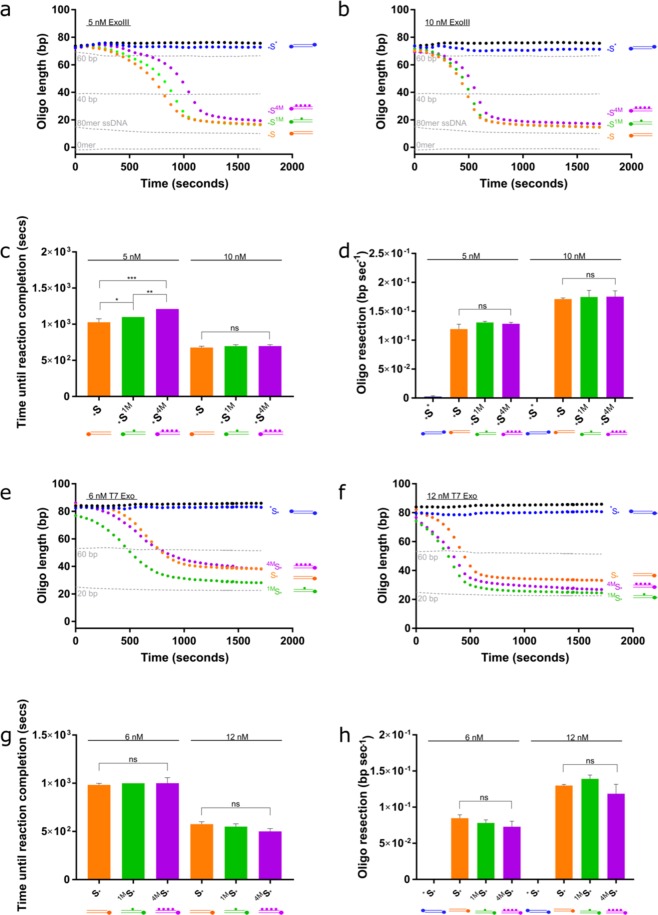
Increased methylcytosine content delays ExoIII-mediated resection; but does not affect the rate of resection of either ExoIII or T7 Exonuclease. (a) 5 nM and (b) 10 nM ExoIII was added to a non-methylated substrate, a substrate containing one methylated cytosine, and a substrate containing four methylated cytosines (*S, *S1M and *S4M, respectively). Standard curve is represented by the grey dotted lines. (c) Time (seconds) until the ExoIII reaction reaches completion on the methylated and unmethylated substrates based on the point at which the graphs plateau in (a) and (b). (d) Calculated resection rate of ExoIII based on maximum gradients in (a) and (b). (e) 6 nM and (f) 12 nM T7 Exo on non-methylated and differentially methylated substrates. Standard curve is represented by the grey dotted lines. (g) Time (seconds) until the T7 Exo reaction reaches completion on the methylated and unmethylated substrates based on point at which the graphs plateau in (e) and (f). (h) Calculated resection rate of ExoIII. Error bars represent SEM; n = 3 in all cases; *p < 0.05, **p < 0.01, ***p < 0.001.

