Figure 1.
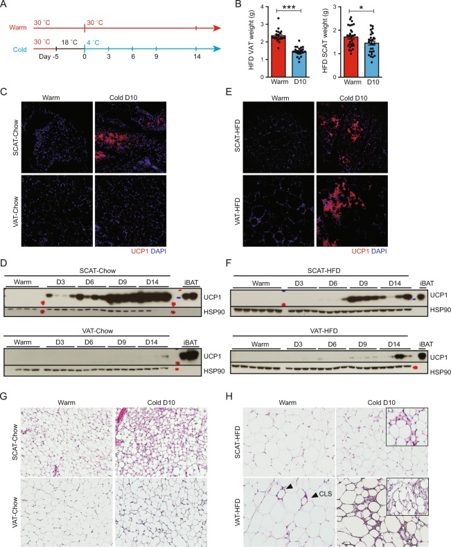
Cold exposure induces weight loss and browning in obese mice. C57BL/6 mice on either normal chow (10% kcal fat) or HFD (60% kcal fat) were maintained at thermoneutrality (30°C, Warm) or exposed to 4°C (Cold) after adaptation at 18°C. (A) The design of the experiment. (B) VAT and SCAT weights after 10-days 4°C cold exposure (N ≥ 18). *P < 0.05, ***P < 0.001 by Student’s t-test. Data are shown as mean ± SEM. (C) Representative IF for UCP1 (red) in lean SCAT and VAT. (D) Western blot analysis for UCP1 expression in lean mice maintained at thermoneutrality or exposed to 4°C for an indicated time as in Fig. 1A. HSP90 serves as loading control. (E) Representative IF for UCP1 (red) in HFD-fed SCAT and VAT. DAPI (blue) stains cell nuclei. (F) Western blot analysis for UCP1 expression in obese mice maintained at thermoneutrality or exposed to 4°C for an indicated time as in Fig. 1A. HSP90 serves as loading control. (G) H&E staining in lean SCAT and VAT. (H) H&E staining in HFD-fed SCAT and VAT. CLS in Warm VAT are indicated by triangles. Inlets highlight separate areas within the same section exhibiting multilocular beige adipocytes.

