Fig. 1.
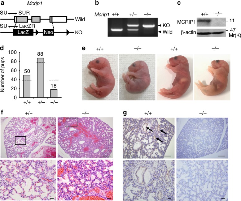
Mcrip1-KO mice die at the neonatal stage due to respiratory failure. a The targeting strategy for Mcrip1-KO mouse generation. Positions of the PCR primers (SU, SUR, and LacZR; for details see methods) that were used for genotyping are indicated. b PCR analysis of genomic DNA isolated from Mcrip1 wild-type (+/+), heterozygous (+/−), or homozygous (−/−) KO mice. c Immunoblot analysis of MCRIP1 expression in MEFs isolated from Mcrip1+/+ or Mcrip1−/− mice. β-actin, loading control. d Genotype distribution of mice from a pool of 156 recorded pups that were still alive at P7. The bar graph indicates the observed number of pups; the dashed line indicates the expected number of pups according to Mendel’s law. e Gross morphology of Mcrip1+/+ and Mcrip1−/− newborn mice. f Hematoxylin and eosin staining of lungs from Mcrip1+/+ or Mcrip1−/− mice at P0. The areas in the small squares in the upper panels were enlarged and are shown below. g Immunohistochemical analysis of MCRIP1 expression in Mcrip1+/+ and Mcrip1−/− lungs. Lung tissues isolated from the neonates were sectioned and stained with an anti-MCRIP1 antibody. Arrows indicate the MCRIP1-expressing epithelial layer in Mcrip1+/+ lungs. f, g Scale bars, 100 μm (upper), 50 μm (lower)

